

If you’ve ever been curious about the world of medical imaging, you’ve likely encountered the term "sonography." While many people immediately think of baby photos, diagnostic medical sonography is a complex, high-tech field that serves as a cornerstone of modern healthcare.
To help you understand what this career actually looks like, we’ve broken down the most common questions using insights directly from our lab and faculty.
1. What exactly does a sonographer do?
Sonographers are frontline medical professionals trained to capture and analyze images in real-time. Because ultrasound is non-invasive and doesn't use radiation, it is often the first step to rule out disease or identify pathologies.
Beyond just taking pictures, you are responsible for patient care, communicating with physicians (like radiologists, obstetricians and cardiologists), and alerting these professionals to urgent findings.
2. What kind of equipment will I use?
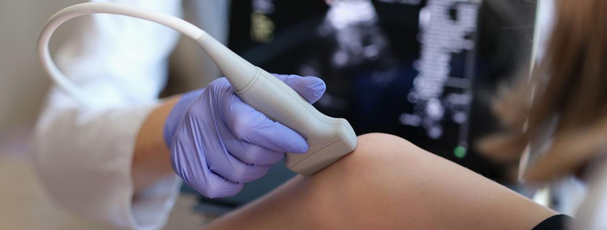
The "camera" of an ultrasound machine is called a transducer. It’s a sophisticated tool that converts electrical signals into sound waves and then translates the "echoes" back into images.
One of the most interesting parts of the job is that "one size does not fit all." You have to choose the right transducer based on the patient’s age, body mass index, and the specific organ you are viewing. It requires a high degree of "psycho-motor skill", which means excellent hand-eye coordination to manipulate the probe and find the perfect view.
3. Is it just about looking at black-and-white images?
While the standard "2D B-mode" (the grey-scale images) is the foundation, modern sonography is incredibly high-tech. Today’s units have over 50 different control features to optimize image quality.
As a student, you'll learn ‘knobology’, the art of using the instrumentation panel to toggle between different modes, such as:
- Color Doppler: To assess blood flow.
- M-mode: To track the motion of a beating heart.
- 3D Ultrasound: Used for detailed obstetric and cardiac exams.
Getting these settings right is critical; a poorly optimized image can lead to a false diagnosis or a missed pathology, which is why your technical training is so vital.
4. Who are the patients I will be helping?
One of the most rewarding aspects of sonography is the variety. You aren't tucked away in a dark room; you are interacting with people at every stage of life. You might monitor a tiny embryo in the first trimester, scan a premature neonate in the NICU, or help an elderly patient with vascular concerns. Because it is painless and safe, it is the preferred imaging choice for almost everyone—even in veterinary medicine!
5. How do I get started?
The path to becoming a sonographer requires a blend of academic knowledge and hands-on skills. You’ll need to master anatomy, understand pathology and disease processes, understand the physics of sound, and the clinical judgment required to help doctors save lives. Sonography Canada sets the standards for the new technologists and educational institutions need to teach to these standards.
Explore Mohawk College’s sonography programs and learn which is right for you:
- Ultrasonography - Medical Radiation Sciences (750)
- Medical Radiation Sciences (729)
- Diagnostic Cardiac Sonography (717)